When Should You See a Vascular Surgeon for Pelvic Pain?
Most women with pelvic pain start with a gynaecologist, and rightly so. But when should a vascular surgeon be added to the picture? Here is a clear, practical guide to recognising vascular pelvic pain and understanding the referral and consultation pathway.
For most women, the first port of call for pelvic pain is understandably a gynaecologist. Gynaecologists are trained to rule out and treat the most common causes of pelvic pain, including fibroids, ovarian cysts, pelvic infections, adenomyosis, and endometriosis. However, a meaningful subset of women continue to experience chronic pelvic pain even after a thorough gynaecological work-up returns normal or inconclusive results. It is precisely in this group that a vascular cause, most commonly pelvic congestion syndrome, deserves serious consideration. This article, prepared with input from the clinical practice of Dr. Pritee Sharma, Consultant Vascular and Endovascular Surgeon at Renova Century Hospital, Banjara Hills, Hyderabad, is designed to help patients and referring doctors recognise the signs that point toward a vascular evaluation, and to explain in detail what that evaluation involves.
Why vascular causes of pelvic pain are often missed
Pelvic congestion syndrome remains under-diagnosed for several structural reasons within the healthcare system. First, awareness of the condition among general practitioners and even some specialists remains limited compared to more familiar diagnoses like endometriosis or fibroids. Second, the standard first-line pelvic ultrasound performed in most diagnostic centres is not specifically optimised to detect pelvic venous reflux, meaning the report may come back as normal even when significant venous congestion is present. Third, because the condition sits at the intersection of gynaecology and vascular medicine, patients can sometimes be shuttled between departments without a single specialist taking ownership of the venous possibility. Understanding these gaps is the first step toward knowing when to actively seek a vascular opinion rather than waiting for one to be offered.
Signs and situations that should prompt a vascular consultation
There are several fairly specific clinical scenarios in which a vascular surgeon's input becomes particularly valuable. Recognising these can save patients years of unresolved suffering.
- Chronic pelvic pain lasting more than six months that has not been clearly explained despite gynaecological examination and standard ultrasound.
- Pelvic pain that clearly worsens through the day and improves significantly with rest and leg elevation.
- Pelvic heaviness or dragging discomfort that intensifies after long periods of standing, long commutes, or physically demanding jobs that involve prolonged standing.
- Pain during or persisting for hours after intercourse, particularly a dull post-coital ache rather than sharp momentary pain.
- Visible varicose veins around the vulva, groin, inner thigh, or buttock, especially if they appeared or worsened during or after pregnancy.
- A history of one or more pregnancies, since pregnancy increases pelvic venous pressure and is a recognised risk factor for pelvic congestion syndrome.
- A previous laparoscopy that found little or no endometriosis, yet pain has persisted or only partially improved.
- Unexplained lower back pain that seems to correlate with the pelvic pain pattern rather than a clear musculoskeletal cause.
- Known varicose veins in the legs with an unusual distribution, particularly involving the inner thigh or vulval region, which may suggest a pelvic source of reflux.
What a vascular surgeon adds that a general work-up may not
A vascular surgeon experienced in pelvic venous disease approaches pelvic pain from a different anatomical lens than a gynaecologist. Where a gynaecologist focuses primarily on the uterus, ovaries, tubes, and their surrounding tissue for structural or hormonal disease, a vascular specialist focuses on the venous drainage system of the entire pelvis, including the ovarian veins, internal iliac veins, and their connections to the vulval, thigh, and leg venous systems. This includes specifically assessing for reflux, dilation, and any anatomical compression syndromes such as nutcracker syndrome or May-Thurner syndrome that can silently drive pelvic venous congestion.
This distinct focus means that a vascular consultation is not a duplication of gynaecological care but a complementary one, filling a diagnostic gap that standard pelvic evaluation is not designed to address. In many cases, the two specialists end up working together, with the gynaecologist managing hormonal or structural gynaecological issues while the vascular surgeon manages the venous component, and both communicating to ensure the patient receives coordinated, non-conflicting advice.
What to bring to your first vascular consultation
Coming prepared to your first vascular consultation can make the visit considerably more productive and can help the specialist reach a working diagnosis more quickly. It is helpful to bring a written or mental timeline of when your pain started, how it has changed, and any clear triggers or relieving factors you have noticed, particularly relating to posture, time of day, and menstrual cycle. It is also valuable to bring copies of all previous pelvic ultrasound reports, any CT or MRI scans and their reports, records of any laparoscopy performed along with the operative notes if available, a list of treatments already tried including hormonal medication and their effect on your pain, and a note of any visible vein changes you have noticed on your body, ideally with photographs if the changes are subtle or intermittent.
- Timeline of pain onset, progression, and pattern through the day and menstrual cycle
- All prior pelvic ultrasound, CT, and MRI reports and images, preferably on a disc or accessible digital file
- Laparoscopy operative notes and histopathology reports if a laparoscopy has been performed
- A list of medications and treatments already tried, including their effect
- Photographs of any visible varicose veins around the vulva, thigh, or leg, if present
- A record of pregnancies, deliveries, and any pelvic surgeries
- Any family history of varicose veins or venous disease
What happens at the first visit
The first vascular consultation typically begins with a detailed history focusing on the character, timing, and triggers of the pain, as well as a review of prior investigations and treatments. This is followed by a focused physical examination, which includes looking specifically for varicose veins around the vulva, groin, inner thigh, and leg, since their presence and distribution can offer important clues about an underlying pelvic venous source. The abdomen and lower back are also examined to help rule out other contributing causes.
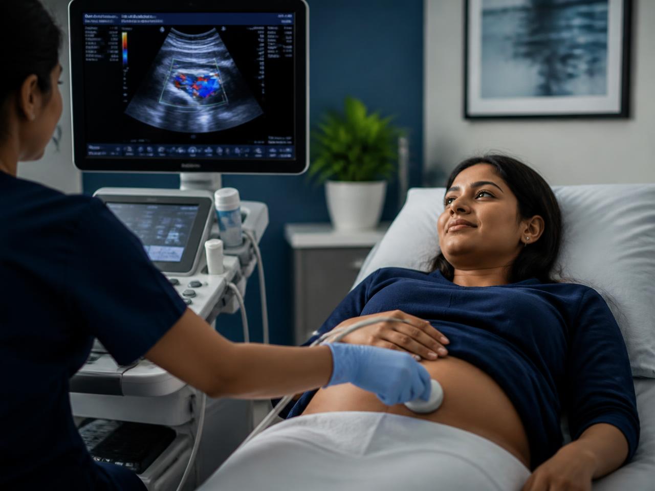
Based on this initial assessment, the vascular surgeon will usually recommend a dedicated pelvic venous ultrasound, sometimes combined with or following a transvaginal ultrasound if this has not already been done with attention to venous flow. Depending on the findings and the level of clinical suspicion, further imaging such as CT venography or MR venography of the pelvis may be advised to map the ovarian and iliac veins in detail and to check for any anatomical compression. Throughout this process, the approach is stepwise, moving from simpler, non-invasive tests toward more detailed imaging only as needed, rather than jumping straight to invasive testing.
The imaging pathway explained step by step
Understanding the logic of the imaging pathway can help reduce anxiety about the process and clarify why certain tests are recommended before others. The pathway generally proceeds in the following order, although the exact sequence can be adjusted based on individual findings.
- Step one, Doppler-assisted pelvic and transvaginal ultrasound: A non-invasive first-line test that looks at the size of the pelvic veins and the direction and speed of blood flow within them, particularly with a Valsalva manoeuvre, which increases abdominal pressure and can reveal reflux that is otherwise not obvious.
- Step two, CT or MR venography: A more detailed cross-sectional scan that maps the ovarian veins, internal iliac veins, and their branches in three dimensions, and can identify anatomical compressions such as nutcracker or May-Thurner syndrome that may be driving the congestion.
- Step three, catheter-based venography: An image-guided procedure performed under local anaesthesia with sedation, where a catheter is passed into the relevant pelvic veins through a small puncture, usually in the neck or groin, and contrast dye is injected under X-ray guidance to directly confirm reflux and vein dilation. This step is both diagnostic and, when confirmed, therapeutic, since embolization can often be performed in the same sitting.
Ovarian vein embolization: what the procedure involves
Once pelvic congestion syndrome is confirmed through catheter venography, the standard treatment is ovarian vein embolization, sometimes extended to the internal iliac veins if they are also found to be refluxing. The procedure is performed by an interventional or vascular specialist in a catheterisation laboratory, under local anaesthesia with light sedation, meaning the patient remains comfortable but does not require general anaesthesia. A catheter is guided through a small puncture in the neck or groin vein into the affected ovarian vein under continuous X-ray guidance. Once the abnormal vein is confirmed, it is blocked using a combination of coils, a sclerosant foam, or a vascular plug, redirecting blood flow away from the congested pathway and toward normal venous channels.
The procedure typically takes between forty-five minutes and a couple of hours depending on how many veins need to be treated, and most patients are observed for a few hours to overnight before discharge. Recovery is generally quick, with most women resuming light activities within a couple of days and full normal activity within a week to ten days, though some residual soreness at the puncture site and mild pelvic discomfort in the first few days is common and expected as the treated veins settle.
What results to expect and how success is measured
Most women who undergo ovarian vein embolization for confirmed pelvic congestion syndrome experience a significant reduction in pelvic heaviness and aching pain over the weeks following the procedure, though the timeline for full improvement varies from person to person and some women notice gradual rather than immediate relief. Improvement is generally assessed through the patient's own reported pain scores and functional improvement, such as ability to stand for longer periods or resume normal activity without the characteristic evening worsening of pain, rather than through repeat imaging alone, although follow-up ultrasound is often performed to confirm the treated veins remain closed.
It is worth setting realistic expectations that, as with any treatment for a chronic condition, a small proportion of women may have partial rather than complete relief, particularly if there is a coexisting contributor to their pain such as endometriosis, in which case addressing that additional factor becomes important for full resolution. This is one more reason why a thorough initial evaluation, considering both gynaecological and vascular contributors, gives the best chance of a satisfying long-term outcome.
Indicative costs of the vascular pathway in Hyderabad
As a general and indicative guide, costs in Hyderabad for the vascular evaluation and treatment pathway can be broadly summarised as follows, though individual quotations will vary by hospital and case complexity. An initial vascular consultation typically costs in the range of INR 800 to 1,500. A dedicated pelvic venous ultrasound may cost approximately INR 2,000 to 5,000. CT or MR venography generally ranges from about INR 8,000 to 20,000 depending on the scan type and facility. Diagnostic catheter venography combined with therapeutic ovarian vein embolization, inclusive of hospital stay, typically falls in the range of INR 1,00,000 to 2,50,000, depending on the number of veins treated and the specific embolic materials used. Patients are strongly advised to request a personalised written estimate from the treating hospital, since insurance coverage and package inclusions vary considerably.
How this fits with leg varicose vein care
Many women who eventually receive a pelvic congestion diagnosis also have some degree of leg varicose veins, and the two are sometimes connected through shared venous drainage pathways between the pelvis and the leg. When a vascular surgeon evaluates a patient for pelvic pain, it is common practice to also assess the leg veins, since treating visible leg varicosities without addressing an underlying pelvic source can lead to early recurrence after leg vein treatment. Readers who have already been diagnosed with or treated for leg varicose veins, and who separately notice pelvic heaviness or vulval vein changes, may benefit from mentioning both issues during consultation so they can be evaluated together. More detail on modern leg vein treatment options is available on the varicose veins treatment page.
The role of a dedicated pelvic vein clinic
Because pelvic congestion syndrome sits at the crossroads of gynaecology and vascular medicine, and because so many women have already been through a long and sometimes discouraging diagnostic journey before reaching a correct diagnosis, a dedicated clinical pathway focused specifically on pelvic venous disorders can make a substantial difference. The Pelvic Vein Clinic has been set up with exactly this purpose, to provide a focused, efficient pathway for women with suspected pelvic congestion syndrome, from the initial history and examination through to targeted imaging and, where appropriate, minimally invasive treatment, all coordinated in one place rather than scattered across multiple unrelated appointments.
Dr. Pritee Sharma's work in this specific area has also received public attention, including a feature in the Times of India highlighting pelvic congestion syndrome as an under-recognised cause of chronic pelvic pain among Indian women, reflecting a broader effort to bring this condition into mainstream awareness so that fewer women spend years searching for an explanation for their pain.
Talking to your gynaecologist about a vascular referral
If you suspect your symptoms may fit the vascular pattern described in this article, it is entirely reasonable to raise this possibility with your gynaecologist and ask specifically whether a pelvic venous ultrasound or a vascular referral would be appropriate, particularly if your standard work-up has been inconclusive. Most gynaecologists are receptive to collaborative care and will welcome a vascular opinion when gynaecological causes have been reasonably excluded or only partially explain the symptoms. If you feel your concerns are not being taken seriously, or if you would simply prefer to start directly with a vascular evaluation given a strong symptom match, you are equally welcome to book a consultation independently.
Red flags requiring urgent rather than routine evaluation
While pelvic congestion syndrome and most causes of chronic pelvic pain are not medical emergencies, certain warning signs should prompt urgent care rather than a scheduled outpatient visit. These include sudden, severe pelvic pain, especially with fever or fainting, which may indicate an acute gynaecological emergency such as ovarian torsion or a ruptured cyst; heavy or unusual vaginal bleeding accompanying severe pain; and a swollen, warm, or tender leg, which could indicate deep vein thrombosis and needs prompt assessment rather than being assumed to be simple venous congestion. Any of these symptoms warrant immediate medical attention at an emergency facility.
Preparing questions for your consultation
Patients often find it useful to prepare a short list of questions in advance to make the most of a specialist consultation, particularly when seeing a new type of specialist for the first time. Useful questions to consider asking include whether your pain pattern is consistent with pelvic venous congestion, what specific imaging is being recommended and why, what the imaging is expected to show or rule out, what the treatment options would be if congestion is confirmed, what the procedure involves and what recovery would look like, and what the realistic likelihood of significant pain improvement would be given your specific findings.
- Does my pain pattern fit with pelvic venous congestion based on what you have heard so far?
- What imaging do you recommend next, and what specifically will it help confirm or rule out?
- If pelvic congestion is confirmed, what would the treatment involve, and is embolization suitable for me?
- What can I expect during recovery, and when can I return to normal activity?
- Should I also be evaluated for endometriosis or other gynaecological causes alongside this?
- What is the realistic likelihood of meaningful pain improvement in a case like mine?
About Dr. Pritee Sharma
Dr. Pritee Sharma is a DNB Gold Medalist and Consultant Vascular and Endovascular Surgeon with more than 21 years of clinical experience, currently practising at Renova Century Hospital in Banjara Hills, Hyderabad. Her clinical interests include the diagnosis and minimally invasive treatment of pelvic congestion syndrome, varicose veins, and a broad range of venous and arterial conditions. Her efforts to raise awareness of pelvic congestion syndrome, including recognition through a feature in the Times of India, reflect her ongoing commitment to improving early recognition of this often-overlooked condition among Indian women. To learn more about her training and clinical approach, visit the about page, and if the concerns raised in this article resonate with your own experience, the most useful next step is to book a consultation through the contact page so a personalised assessment and imaging plan can be arranged.
Key takeaways
- A vascular surgeon's opinion is worth seeking when pelvic pain is postural, worsens through the day, and has not been explained by standard gynaecological evaluation.
- Vascular pelvic pain evaluation is complementary to, not a replacement for, gynaecological care.
- The imaging pathway moves stepwise from Doppler-assisted ultrasound to CT or MR venography and, if needed, catheter venography.
- Ovarian vein embolization is a minimally invasive, image-guided treatment that can often be performed in the same sitting as the diagnostic venogram.
- Bringing prior reports, a clear symptom timeline, and photographs of any visible vein changes makes the first vascular consultation far more productive.
- A dedicated pelvic vein clinic pathway can shorten the diagnostic journey for women who have been searching for answers for months or years.
- Varicose Veins Treatment in Hyderabad
- Peripheral Artery Disease Treatment
- Diabetic Foot Care Specialist
- DVT Treatment in Hyderabad
- Angioplasty for Blocked Leg Arteries
- Carotid Surgery — Stroke Prevention
- Venous Stenting for Chronic Leg Swelling
- AV Fistula Creation for Dialysis
- EVAR — Endovascular Aneurysm Repair
- TEVAR — Thoracic Aortic Repair
- About Dr. Pritee Sharma
- Vascular surgery FAQs