What Is Pelvic Congestion Syndrome? A Complete Guide
A complete, medically grounded guide to pelvic congestion syndrome, covering causes, symptoms, diagnostic imaging, ovarian vein embolization, recovery, costs in Hyderabad and when to seek specialist help.
Understanding Pelvic Congestion Syndrome
Pelvic congestion syndrome is a chronic condition in which faulty, dilated veins within the pelvis cause persistent pelvic pain, heaviness and discomfort, most often in women who have been pregnant one or more times. It arises from a mechanism very similar to varicose veins in the legs, except that the affected veins lie deep within the pelvis, around the ovaries, uterus and broad ligament, and are therefore invisible on ordinary physical examination. Because the pain is real but the cause is hidden, many women spend years moving between gynaecologists, gastroenterologists, urologists and even psychiatrists before the correct diagnosis is reached.
In a busy clinical practice in Hyderabad, it is common to see women in their late twenties to forties who describe a dull, dragging ache low in the pelvis that has been present for months or years, often dismissed as a normal part of having had children or as stress-related discomfort. This guide is written to give a complete, honest and medically grounded picture of what pelvic congestion syndrome actually is, why it happens, how it is diagnosed and what can be done about it, including modern minimally invasive treatment.
The Venous Anatomy Behind the Problem
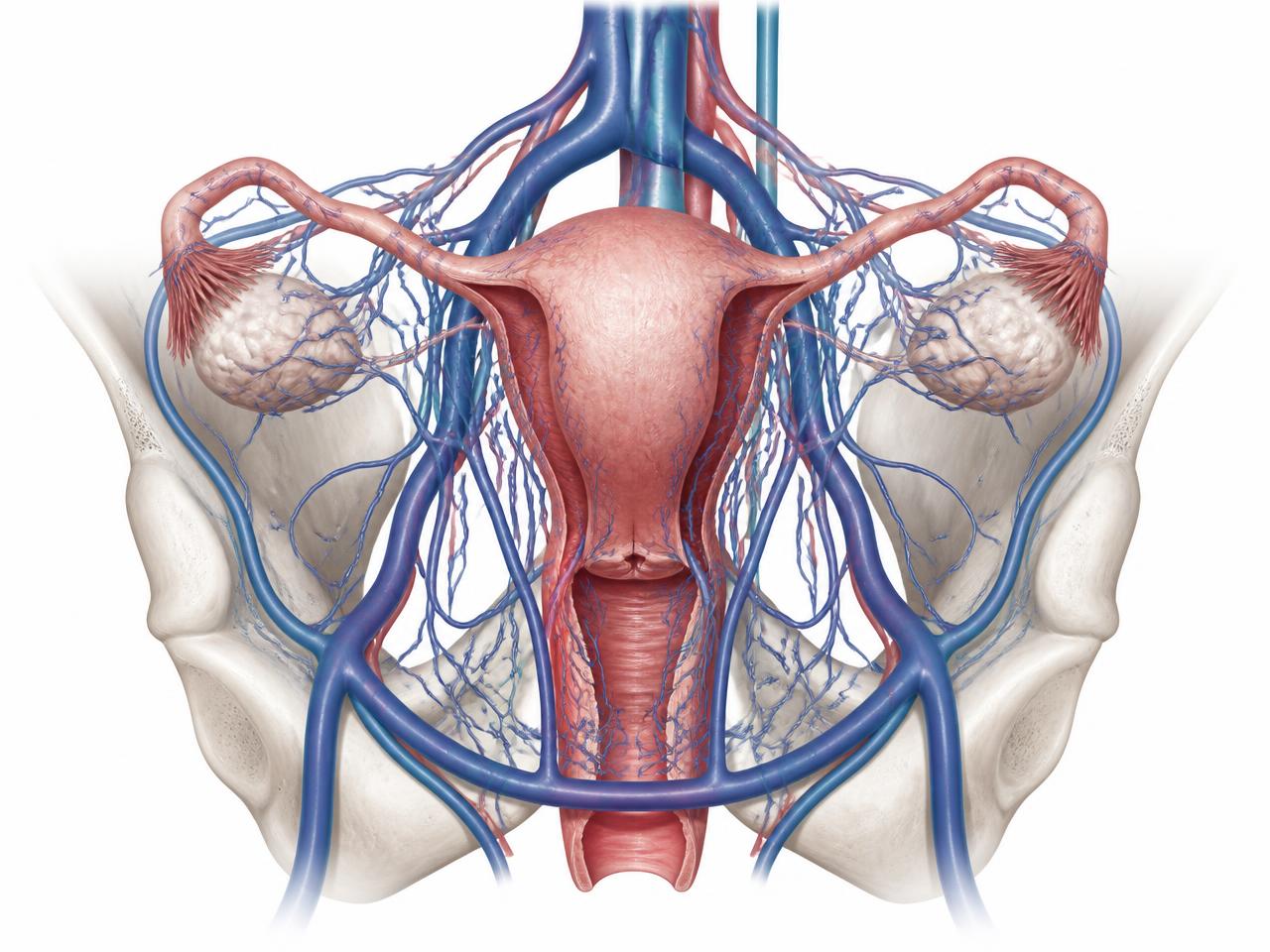
To understand pelvic congestion syndrome, it helps to understand how blood normally leaves the pelvic organs. Blood flows into the uterus and ovaries through arteries, and then must be carried back to the heart through a network of veins, principally the ovarian veins and the internal iliac veins and their tributaries. The right ovarian vein usually drains directly into the inferior vena cava at a relatively steep angle, while the left ovarian vein typically drains into the left renal vein at a right angle. This anatomical difference is one reason why the left ovarian vein is more frequently involved in pelvic congestion syndrome than the right.
Like the veins in the legs, ovarian and internal iliac veins contain one-way valves whose job is to prevent blood from flowing backward under the pull of gravity or increased abdominal pressure. When these valves become weak, stretched or incompetent, blood pools within the vein instead of moving efficiently toward the heart. Over time this pooling causes the vein to dilate further, worsening valve function in a self-reinforcing cycle. The dilated, sluggish veins around the ovary and uterus form a cluster often described on imaging as a pelvic venous plexus, and it is this congested, high-pressure network of veins that produces the characteristic dragging pain of the syndrome.
It is worth noting that not every woman with dilated pelvic veins on a scan has symptomatic pelvic congestion syndrome. Some degree of pelvic venous dilation can be an incidental finding, and the diagnosis is properly made only when imaging findings are combined with a compatible clinical picture and after other causes of pelvic pain have been reasonably excluded.
Why Pregnancy and Multiple Childbirths Matter
Pregnancy is one of the strongest risk factors for pelvic congestion syndrome, and the risk appears to increase with each successive pregnancy. During pregnancy, blood volume increases substantially, hormonal changes relax and soften vein walls, and the growing uterus places direct mechanical pressure on the pelvic veins, all of which combine to stretch the veins and strain their valves. In many women, veins that were stretched during pregnancy do not fully return to their pre-pregnancy calibre afterward, leaving behind a degree of permanent dilation and valve weakness that can slowly progress over subsequent years.
This explains why pelvic congestion syndrome is uncommon in women who have never been pregnant and becomes progressively more frequent with increasing numbers of pregnancies. It also explains the typical age group affected, since most women are diagnosed in their reproductive years, generally between the mid-twenties and mid-forties, at a time when the cumulative effect of one or more pregnancies has had time to manifest as symptomatic venous disease.
- Multiple pregnancies (multiparity), which cumulatively stretch pelvic veins
- Family history of varicose veins or venous insufficiency
- Hormonal factors, since oestrogen is known to soften and dilate vein walls
- Occupations or lifestyles involving prolonged standing
- Retroverted uterus or other anatomical variations that alter venous drainage
- Obesity, which increases intra-abdominal pressure
- Nutcracker phenomenon, where the left renal vein is compressed and backs up pressure into the left ovarian vein
Typical Symptoms Women Describe
The hallmark symptom of pelvic congestion syndrome is a chronic, dull, aching or heavy pain low in the pelvis that has usually been present for at least six months, which is the general threshold used to define chronic pelvic pain in clinical practice. Unlike the sharp, colicky pain of conditions such as ovarian cysts or kidney stones, the pain of pelvic congestion syndrome tends to be a persistent background ache that fluctuates in intensity rather than coming and going in dramatic spasms.
Several features tend to be characteristic, even though no single symptom is unique to this condition. Pain typically worsens as the day progresses, particularly after long periods of standing or sitting, and tends to ease somewhat with lying down and elevating the legs, mirroring the pattern seen in leg varicose veins. Pain often intensifies premenstrually and during or after sexual intercourse, a symptom called deep dyspareunia that many women find embarrassing to discuss but which is an important diagnostic clue. Pain may also worsen with prolonged travel, heavy lifting or straining.
- Chronic dull, aching or heavy pelvic pain lasting more than six months
- Pain that worsens with standing and improves with lying flat
- Deep pain during or after sexual intercourse
- Worsening of pain before or during menstruation
- Visible varicose veins around the vulva, inner thigh or buttock
- A sensation of heaviness or fullness in the pelvis
- Low backache without an obvious spinal cause
- Associated leg varicose veins in a proportion of women
Many women also notice visible varicose veins in the vulval region, inner thighs or buttocks, which occur because the congested pelvic veins communicate with superficial veins outside the pelvis. When a woman presents with both unexplained chronic pelvic pain and unusual varicose veins in these locations, pelvic congestion syndrome should be actively considered rather than treated as an afterthought.
How the Diagnosis Is Reached
Diagnosing pelvic congestion syndrome is a process of careful clinical reasoning combined with targeted imaging, since there is no single blood test that confirms the condition. The first step is always a detailed history and physical examination, during which the treating doctor will ask about the pattern, timing and triggers of the pain, obstetric history, menstrual history and any associated symptoms such as urinary or bowel complaints that might point toward an alternative diagnosis.
Doppler ultrasound of the pelvis, performed both transabdominally and transvaginally, is usually the first imaging test used. Transvaginal ultrasound with Doppler allows detailed visualisation of the ovarian and pelvic veins, showing dilated, tortuous veins with slow or reversed blood flow, which are the classic sonographic signs of pelvic venous incompetence. This test is widely available, does not involve radiation and can often be performed on the same day as the initial consultation.
When ultrasound findings are suggestive but not entirely conclusive, or when a more detailed anatomical map is required before planning treatment, cross-sectional imaging such as CT venography or MR venography is used. These techniques give a three-dimensional view of the ovarian veins, internal iliac veins and their tributaries, and can also help exclude other pelvic pathology such as fibroids, ovarian cysts, endometriosis or adenomyosis that may be contributing to or mimicking the pain. MR venography has the advantage of avoiding ionising radiation and is particularly useful in younger women.
The definitive, gold-standard investigation remains catheter-based pelvic venography, performed at the same sitting as ovarian vein embolization if treatment is planned. In this procedure, a thin catheter is guided under X-ray guidance into the ovarian and internal iliac veins, and contrast dye is injected to directly visualise the abnormal, refluxing veins in real time. Because it is minimally invasive and combines diagnosis with the option of immediate treatment, catheter venography is generally reserved for women in whom non-invasive imaging has already raised a strong suspicion of pelvic congestion syndrome.
- Clinical history and pelvic examination
- Transabdominal and transvaginal Doppler ultrasound
- CT venography for detailed anatomical mapping
- MR venography, particularly useful in younger women
- Catheter pelvic venography, the gold-standard confirmatory and often therapeutic test
Ruling Out Other Causes of Pelvic Pain
Chronic pelvic pain in women can arise from gynaecological, gastrointestinal, urological and musculoskeletal causes, and pelvic congestion syndrome is only one entry on a fairly long list. A thorough evaluation, usually involving both a gynaecologist and a vascular surgeon, is important before treatment is planned, both to avoid missing a more urgent diagnosis and to identify situations where more than one condition is contributing to the pain simultaneously.
- Endometriosis, which can cause cyclical or constant pelvic pain
- Uterine fibroids or adenomyosis
- Ovarian cysts or chronic pelvic inflammatory disease
- Irritable bowel syndrome or other gastrointestinal disorders
- Interstitial cystitis or other bladder-related pain syndromes
- Musculoskeletal causes such as pelvic floor dysfunction
It is entirely possible, and in fact common, for a woman to have both pelvic congestion syndrome and a coexisting gynaecological condition such as mild endometriosis. In such cases, treating only one condition may lead to incomplete relief, which is why a collaborative approach between specialities generally gives the best outcome.
Conservative and Medical Treatment Options
For women with mild symptoms, or as a first step before considering intervention, several conservative measures can help reduce discomfort. These include wearing supportive compression garments designed for the pelvic region, avoiding prolonged standing where possible, maintaining a healthy body weight, and using simple pain relief medication during flare-ups. Some doctors also prescribe hormonal medications that reduce ovarian venous congestion, though these are generally used as a temporary measure rather than a definitive solution, since symptoms often return once the medication is stopped.
Conservative treatment can meaningfully improve quality of life for women with milder disease, but it does not correct the underlying valve incompetence, and women with moderate to severe symptoms that interfere with daily activities, intimacy or work usually require a more definitive procedure to achieve lasting relief.
Ovarian Vein Embolization: The Modern Treatment
Ovarian vein embolization is currently the most widely used and best-established minimally invasive treatment for pelvic congestion syndrome. It is performed by an interventional or vascular surgeon under local anaesthesia with sedation, avoiding the need for general anaesthesia or a surgical incision. A small catheter, usually introduced through a vein in the neck or groin, is guided under X-ray guidance into the affected ovarian vein or internal iliac vein tributaries.
Once the abnormal vein has been mapped using contrast venography, small coils, a sclerosing agent, or a combination of both are deployed within the vein to permanently close it off. Blocking the incompetent vein prevents blood from pooling in the congested pelvic venous plexus, allowing pressure within the pelvic veins to normalise over the following weeks. Because both ovarian veins and sometimes branches of the internal iliac veins can be involved, a thorough procedure may treat more than one vein in the same sitting.
The procedure typically takes between forty-five minutes and two hours depending on the complexity of the venous anatomy, and most women can go home the same day or after an overnight stay for observation. Recovery is generally quick compared to any form of open or laparoscopic surgery, with most women returning to light activity within two to three days and to full normal activity within one to two weeks. Some mild pelvic discomfort or a dull ache in the days following the procedure is common and expected, as the treated veins settle.
Across published clinical experience worldwide, ovarian vein embolization is generally reported to provide meaningful symptom relief in roughly seventy-five to eighty-five percent of appropriately selected women, with many experiencing a significant reduction in pelvic pain and improvement in quality of life within a few months of the procedure. It is important to set realistic expectations: a smaller proportion of women may have only partial relief, particularly if other contributing conditions such as endometriosis are also present, and a very small number may need a repeat procedure if a previously untreated vein becomes symptomatic later.
- Minimally invasive, performed through a small needle puncture rather than an incision
- Local anaesthesia with sedation, avoiding general anaesthesia in most cases
- Same-day or overnight hospital stay
- Recovery to light activity within two to three days for most women
- Meaningful symptom relief in a majority of appropriately selected patients
Risks and Safety Considerations
Ovarian vein embolization is generally a safe procedure when performed by an experienced vascular or interventional specialist, but like any medical procedure it carries some risk. Possible complications include bruising or discomfort at the catheter entry site, a small risk of the coil migrating from its intended position, transient worsening of pain in the first few days, and a low risk of the vein recanalising or an untreated tributary continuing to cause symptoms. Radiation exposure during the procedure is kept as low as reasonably possible through careful imaging technique, and the overall complication rate reported in the literature is low.
A frank discussion of these risks, along with realistic expected benefits, should form part of every pre-procedure consultation, and women should feel comfortable asking their treating doctor about the specific numbers relevant to their own case and the operator's personal experience with the procedure.
Indicative Costs in Hyderabad
Costs for the diagnostic pathway and treatment of pelvic congestion syndrome in Hyderabad vary depending on the hospital, the complexity of the case, the number of veins treated and whether the patient has health insurance. As a broad and purely indicative guide, an initial vascular consultation may cost in the range of INR 800 to 2,000, a transvaginal Doppler ultrasound typically costs approximately INR 2,000 to 5,000, and cross-sectional venography such as CT or MR venography can range from roughly INR 8,000 to 20,000 depending on the imaging centre and protocol used.
Ovarian vein embolization itself, including hospital stay, catheter equipment, coils or sclerosant, and the procedural fee, generally falls in the range of INR 1,50,000 to 3,50,000 in a good private hospital setting in Hyderabad, though this can vary based on the number of veins embolized and the materials used. These figures are indicative only and patients should obtain a personalised estimate during consultation, since insurance coverage, hospital package inclusions and individual anatomical complexity can all affect the final cost.
When to See a Vascular Surgeon
Women who have experienced pelvic pain for more than six months, especially pain that worsens with standing and improves with lying down, pain during or after intercourse, or visible unusual varicose veins around the vulva or upper thighs, should consider a dedicated vascular evaluation in addition to routine gynaecological review. Early involvement of a vascular surgeon experienced in pelvic venous disease can shorten the diagnostic journey considerably and avoid years of trial-and-error treatment for a condition that has a well-defined, minimally invasive solution.
If you or a loved one is struggling with unexplained pelvic pain that has not responded to standard gynaecological treatment, consider a consultation at the Pelvic Vein Clinic (/pelvic-vein-clinic), where a focused, vein-specific evaluation is offered alongside routine gynaecological and general surgical assessment. Dr. Pritee Sharma's work in this field was featured in a Times of India article on pelvic congestion syndrome, which helped bring wider public and physician awareness to this frequently missed cause of chronic pelvic pain in Indian women.
Readers who also have visible varicose veins in the legs, or a family history of venous disease, may find it useful to read more about lower-limb venous disease on the varicose veins treatment page (/services/varicose-veins-treatment-hyderabad), since pelvic and leg venous insufficiency frequently coexist and share a common underlying tendency toward weak vein walls and valves. More about Dr. Pritee Sharma's training, credentials and approach to vascular care is available on the about page (/about-dr-pritee-sharma), and appointments or queries can be directed through the contact page (/contact).
A Final Word
Pelvic congestion syndrome remains under-recognised, partly because it cannot be seen or felt on a routine physical examination and partly because awareness of pelvic venous disease among both patients and some treating doctors is still growing in India. With the right combination of clinical suspicion, targeted imaging and, where appropriate, minimally invasive ovarian vein embolization, the great majority of affected women can achieve substantial and lasting relief from a condition that too often goes undiagnosed for years.
- Varicose Veins Treatment in Hyderabad
- Peripheral Artery Disease Treatment
- Diabetic Foot Care Specialist
- DVT Treatment in Hyderabad
- Angioplasty for Blocked Leg Arteries
- Carotid Surgery — Stroke Prevention
- Venous Stenting for Chronic Leg Swelling
- AV Fistula Creation for Dialysis
- EVAR — Endovascular Aneurysm Repair
- TEVAR — Thoracic Aortic Repair
- About Dr. Pritee Sharma
- Vascular surgery FAQs