PCS vs Endometriosis: How to Tell the Difference
Chronic pelvic pain in women is very often blamed on endometriosis alone, but pelvic congestion syndrome frequently coexists with it or mimics it. Here is a detailed, practical comparison to help you and your doctors reach the right diagnosis sooner.
Chronic pelvic pain is one of the most common reasons women in Hyderabad, and across India, visit a gynaecologist. It is also one of the most frustrating conditions to diagnose, because so many different organ systems sit crowded together in the pelvis. Two of the most frequently confused causes are endometriosis and pelvic congestion syndrome, commonly shortened to PCS. Both can cause months or years of pain, both can affect fertility and quality of life, and both are still under-recognised by many first-contact doctors. This article is written to help patients understand the differences, the overlaps, and the pathway to an accurate diagnosis, drawing on the clinical experience of Dr. Pritee Sharma, Consultant Vascular and Endovascular Surgeon at Renova Century Hospital, Banjara Hills, Hyderabad.
It is important to say at the outset that this comparison is not meant to replace a proper medical evaluation. Pelvic pain has dozens of possible causes, from ovarian cysts and adenomyosis to irritable bowel syndrome, interstitial cystitis, musculoskeletal strain, and psychological contributors. What this article aims to do is sharpen your awareness of two specific and often overlapping conditions so that you can ask the right questions when you see your doctor, and so that you know when a vascular opinion may be worth seeking alongside a gynaecological one.
What is endometriosis, in simple terms
Endometriosis is a condition in which tissue similar to the lining of the uterus, the endometrium, grows outside the uterine cavity. These deposits can appear on the ovaries, the fallopian tubes, the outer surface of the uterus, the bowel, the bladder, and the peritoneal lining of the pelvis. Because this tissue responds to the hormonal cycle in the same way the uterine lining does, it can bleed internally each month, causing inflammation, scarring, and the formation of adhesions that bind pelvic organs together. Over years, this process can distort pelvic anatomy and create chronic pain, heavy periods, painful intercourse, and, in a significant proportion of women, difficulty conceiving.
Endometriosis is primarily a gynaecological and, in advanced cases, a surgical diagnosis. It is confirmed definitively by laparoscopy, where a surgeon directly visualises endometrial deposits, cysts called endometriomas, and any associated scarring, and can take a biopsy to confirm the tissue type under a microscope.
What is pelvic congestion syndrome, in simple terms
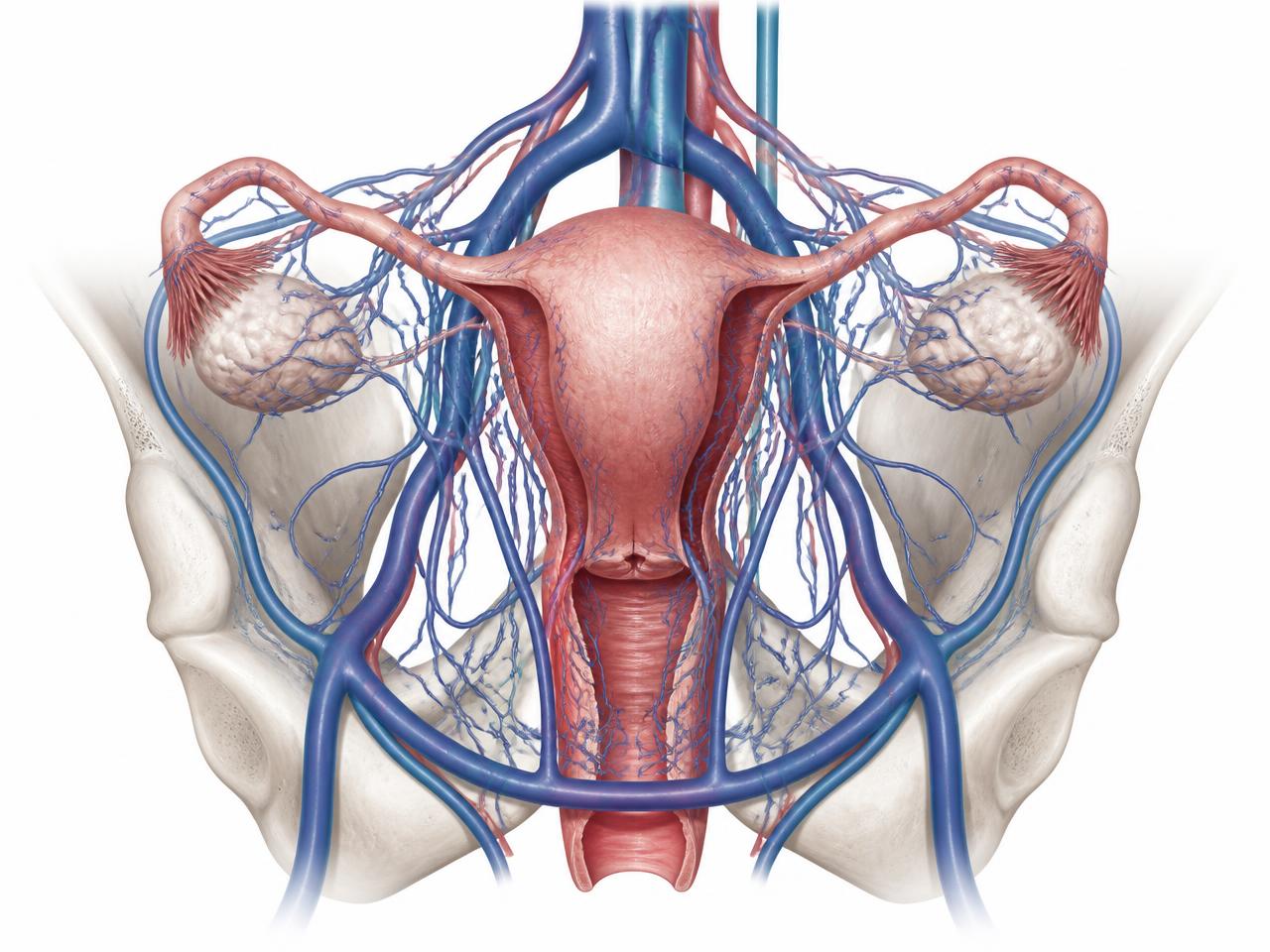
Pelvic congestion syndrome is a vascular condition. It occurs when the ovarian veins, and sometimes the internal iliac veins, fail to carry blood efficiently back toward the heart from the pelvis. The valves inside these veins, which normally act as one-way gates preventing backward flow, become weak or incompetent. Blood pools and flows backward, a phenomenon called venous reflux. Over time this leads to dilated, tortuous veins around the ovary and uterus, similar in principle to varicose veins in the legs but located deep inside the pelvis where they cannot be seen from the outside.
This pooling of blood stretches the vein walls and the surrounding ligaments and tissues, which are richly supplied with pain nerve fibres. The result is a dull, heavy, aching pelvic pain that characteristically worsens through the day, after standing for long periods, after intercourse, and during or just before menstruation. PCS is a diagnosis made primarily through imaging, particularly transvaginal ultrasound with Doppler, and confirmed with CT or MR venography or catheter-based venography, rather than through direct surgical visualisation.
Why these two conditions are so often confused
Both endometriosis and pelvic congestion syndrome typically affect women of reproductive age. Both can cause pain that fluctuates with the menstrual cycle. Both can cause pain during intercourse, known as dyspareunia. Both can be worsened by prolonged standing or physical activity in some patients. Both are frequently missed for years because standard pelvic examinations and basic ultrasound scans can appear entirely normal, especially in early or mild disease. It is not unusual for a woman to consult three or four doctors, undergo a laparoscopy that shows minimal or no endometriosis, and still be left with unexplained pain, only to later be found to have significant pelvic venous congestion on a dedicated vascular scan.
Adding further complexity, the two conditions are not mutually exclusive. Research and clinical experience both suggest that a meaningful proportion of women with confirmed endometriosis also have coexisting pelvic venous insufficiency, and the inflammation from endometriosis may itself contribute to or worsen venous congestion in some patients. This means a woman can genuinely have both conditions contributing to her symptoms at the same time, which is one reason why treating only one may leave residual pain even after apparently successful surgery.
Comparing the pain: character, timing, and triggers
While no single symptom can definitively separate the two conditions without imaging or surgery, there are patterns that experienced clinicians look for. The following comparison summarises typical, though not universal, differences observed in clinical practice.
- Pain quality: Endometriosis pain is often described as sharp, cramping, or stabbing, especially around and during menstruation. PCS pain is more often described as a dull, heavy, dragging ache, sometimes likened to a sensation of fullness or heaviness low in the pelvis.
- Timing through the day: Endometriosis pain does not typically change much with posture or time of day, though it peaks with the menstrual cycle. PCS pain classically worsens as the day progresses, being mildest in the morning after lying down all night and most severe by evening, especially after standing or sitting for long periods.
- Relationship to intercourse: Both conditions can cause dyspareunia, but endometriosis pain during intercourse is often sharp and felt deep with certain positions, particularly deep penetration, sometimes linked to nodules behind the uterus. PCS-related pain during intercourse is often a dull ache that can persist for hours afterward, sometimes described as a post-coital ache rather than pain only during the act.
- Effect of lying down or rest: Endometriosis pain is not reliably relieved by lying flat. PCS pain characteristically improves significantly when the patient lies down and elevates her legs, because gravity assists venous drainage from the congested pelvic veins.
- Associated leg symptoms: Endometriosis alone does not typically cause visible leg vein changes. PCS is frequently associated with vulval varicose veins, or with varicose veins of the inner thigh, buttock, or leg that seem to originate from the pelvis rather than the usual saphenous vein pattern.
- Bowel and bladder symptoms: Endometriosis, especially deep infiltrating disease, can cause painful bowel movements, rectal bleeding during periods, or bladder pain if deposits involve those organs. PCS is less commonly associated with direct bowel or bladder tissue involvement, though bladder pressure symptoms from congested pelvic veins can occur.
- Response to hormonal therapy: Endometriosis pain often responds, at least partially, to hormonal suppression such as combined pills, progestins, or GnRH analogues, because these reduce the hormonal stimulation of ectopic endometrial tissue. PCS may show variable or only partial response to hormonal therapy since the underlying problem is structural and mechanical, related to vein wall and valve weakness.
Overlap symptoms that do not help distinguish the two
Some symptoms are common to both conditions and therefore cannot, on their own, be used to tell them apart. These include general chronic pelvic pain lasting more than six months, pain that fluctuates with the menstrual cycle, fatigue related to chronic pain and poor sleep, lower back pain, and psychological effects such as anxiety or low mood arising from living with unexplained pain for a long time. Because of this overlap, relying purely on symptom pattern without imaging can lead to misdiagnosis in either direction.
The role of imaging in telling them apart
Imaging is where the two conditions truly begin to separate. A standard transabdominal pelvic ultrasound, the kind most commonly ordered as a first test, is often unrevealing for both conditions unless there is an obvious endometrioma or a very severely congested vein plexus. This is why so many women are told their scan is normal despite significant ongoing pain.
A transvaginal ultrasound performed with attention to venous flow, ideally by a radiologist or clinician experienced in looking for pelvic congestion, can reveal dilated, tortuous pelvic veins, often described as measuring more than four to five millimetres in diameter, with slow or reversed flow on Doppler assessment, particularly when the patient performs a Valsalva manoeuvre. This same transvaginal scan, performed with a gynaecological focus, can also identify ovarian endometriomas, thickened or nodular areas suggestive of deep infiltrating endometriosis, and adhesions restricting normal organ mobility.
When pelvic congestion is suspected based on ultrasound findings and clinical history, CT venography or MR venography of the pelvis provides a more detailed three-dimensional map of the ovarian and internal iliac veins, confirming dilation, reflux, and any associated pelvic varices. These cross-sectional scans can also help rule out other causes of pelvic vein compression, such as nutcracker syndrome, where the left renal vein is compressed between the aorta and the superior mesenteric artery, or May-Thurner syndrome, where the left common iliac vein is compressed by the overlying right common iliac artery. Both of these anatomical compressions can be underlying drivers of pelvic venous congestion and are important to identify because they may change the treatment approach.
The gold standard for confirming pelvic venous reflux, and for treating it in the same sitting, is catheter-based venography. In this procedure, performed under local anaesthesia with sedation, a thin catheter is guided through a vein, usually in the neck or groin, into the ovarian and internal iliac veins under X-ray guidance. Contrast dye is injected to directly visualise reflux and vein dilation in real time. If confirmed, the abnormal veins can often be treated in the same procedure through embolization, discussed further below.
For endometriosis, while imaging has improved considerably and specialised MRI protocols can pick up deep infiltrating disease with good accuracy, laparoscopy remains the only way to achieve a fully definitive diagnosis, because it allows direct visualisation and biopsy of suspicious tissue. This is one of the fundamental practical differences between the two conditions: PCS diagnosis and treatment can largely be achieved through minimally invasive image-guided vascular procedures without general anaesthesia or an abdominal incision, whereas endometriosis diagnosis conventionally requires a surgical, usually laparoscopic, approach.
Why laparoscopy alone can miss pelvic congestion
One of the more important clinical lessons from years of managing chronic pelvic pain is that a normal or near-normal diagnostic laparoscopy does not rule out pelvic congestion syndrome. During laparoscopy, the abdomen is inflated with carbon dioxide gas to create space for the surgeon to see and work. This increased pressure inside the abdomen can temporarily compress and empty the very veins that are congested when the patient is upright and at normal pressure, causing them to look deceptively normal or only mildly dilated on the operating table. Surgeons who are not specifically looking for subtle venous changes, or who are focused on ruling out endometriosis, may report the laparoscopy as unremarkable, and the patient is then left without an explanation for her pain.
This is precisely the scenario in which many women eventually find their way, sometimes after years of consultations, to a vascular surgeon experienced in pelvic venous disease. A careful history revisited with attention to postural worsening of pain, combined with a dedicated venous-focused ultrasound and, if needed, CT or MR venography, can finally provide the missing piece of the puzzle.
When both conditions coexist
It bears repeating that endometriosis and pelvic congestion syndrome are not an either-or diagnosis. A woman can have biopsy-proven endometriosis that has been surgically treated, and still have persistent pain because of untreated pelvic venous congestion. Conversely, a woman diagnosed and treated for pelvic congestion may have partial pain relief but continue to experience cyclical, sharp pain suggestive of coexisting endometriosis that has not yet been evaluated. The inflammatory environment created by endometriosis in the pelvis is also thought to contribute to venous congestion in some women, creating a two-way relationship between the conditions rather than a simple coincidence.
Because of this overlap, the most effective care model for women with persistent, unexplained, or incompletely treated pelvic pain is a collaborative one, where gynaecologists and vascular specialists communicate and, where appropriate, co-manage the patient. A woman who has had a laparoscopy with minimal endometriosis found, but who still has significant heaviness, dragging pain, and postural worsening, should be considered for a pelvic venous evaluation rather than being told that nothing more can be done.
A practical symptom checklist to bring to your doctor
The following checklist, framed around the differences described above, can help you organise your history before a consultation, whether with a gynaecologist or a vascular surgeon.
- Does your pain get noticeably worse as the day goes on, and better when you lie down with your legs raised?
- Do you notice visible varicose veins around the vulva, inner thigh, or buttock, especially during or after pregnancy?
- Is the pain more of a dull, heavy ache rather than sharp cramping, most of the time?
- Does the pain persist for hours after intercourse rather than being sharp only during it?
- Have you had a laparoscopy that showed little or no endometriosis, yet your pain has continued?
- Have you had one or more pregnancies, since pelvic venous dilation is more common after pregnancy?
- Do you also have painful, heavy periods with clots, painful bowel movements during periods, or a history of infertility, which would point more toward endometriosis?
Treatment differences worth understanding
Endometriosis treatment typically involves a combination of hormonal suppression to reduce disease activity and pain, and, when indicated, laparoscopic surgery to excise or ablate visible deposits, remove endometriomas, and free up adhesions. In women who have completed childbearing and have severe, refractory disease, more extensive surgery may occasionally be considered.
Pelvic congestion syndrome, once confirmed, is most commonly and effectively treated with ovarian vein embolization, a minimally invasive procedure performed by an interventional or vascular specialist. Through a small puncture, usually in the neck or groin vein, a catheter is used to access the refluxing ovarian and, if needed, internal iliac veins, which are then blocked off using coils, a sclerosant agent, or a plug, redirecting blood flow through healthy pathways and relieving the pooling and pressure responsible for pain. This procedure is typically done as a day-care or short-stay procedure under local anaesthesia with sedation, avoiding the need for general anaesthesia or an abdominal incision, with most women returning to routine activity within a few days.
For women in whom imaging identifies an anatomical compression such as nutcracker or May-Thurner syndrome as the underlying driver of pelvic congestion, additional treatments such as stenting of the compressed vein may be considered alongside or instead of embolization, depending on the specific findings.
Indicative costs in Hyderabad
Costs for the diagnostic and treatment pathway in Hyderabad vary depending on the hospital, the complexity of the case, and whether additional imaging or procedures are required. As a broad and indicative guide only, a dedicated pelvic venous ultrasound may cost in the range of INR 2,000 to 5,000, CT or MR venography may range from approximately INR 8,000 to 20,000 depending on the modality and facility, and diagnostic catheter venography combined with ovarian vein embolization, including hospital stay, typically falls in the range of INR 1,00,000 to 2,50,000, though this can vary based on the number of veins treated and materials used. These figures are general estimates and any individual patient should obtain a personalised quotation after consultation, since insurance coverage, hospital package inclusions, and clinical complexity all affect the final cost.
The value of seeing both a gynaecologist and a vascular surgeon
Given the substantial overlap between these two conditions, women with chronic pelvic pain that has not been clearly explained, or that has persisted despite treatment for one condition, are best served by seeking a second opinion that specifically addresses the vascular possibility. Dr. Pritee Sharma, who has been featured in the Times of India for her work in raising awareness about pelvic congestion syndrome among Indian women, frequently sees patients referred by gynaecologists once endometriosis has been ruled out or only partially explains the pain picture, as well as patients who come directly seeking clarity after years of unexplained symptoms.
If you are dealing with persistent pelvic heaviness, aching, or pain that seems to fit the pattern described here, especially if you also notice visible vein changes around the pelvis, vulva, or upper thigh, you are warmly invited to visit the dedicated Pelvic Vein Clinic at the Pelvic Vein Clinic page to understand the evaluation process and what to expect. For women who already have diagnosed varicose veins in the legs alongside pelvic symptoms, since the two are often related through shared venous pathways, more information is available on the varicose veins treatment page as well.
Related concerns: leg varicose veins and pelvic congestion
It is worth noting that pelvic venous congestion and leg varicose veins can be connected, because blood refluxing from an incompetent ovarian or internal iliac vein can sometimes find its way into the venous drainage of the vulva, groin, and upper thigh, contributing to unusual patterns of varicose veins that do not follow the typical great or small saphenous vein distribution. In such cases, treating the leg veins alone without addressing the underlying pelvic source can lead to early recurrence after treatment, which is why a thorough vascular evaluation looks at the whole venous system rather than an isolated leg segment. Readers wanting to understand leg vein treatment options in more detail can refer to the dedicated page on varicose veins treatment in Hyderabad.
Red flags that need urgent medical attention
While both endometriosis and pelvic congestion syndrome are generally chronic, non-emergency conditions, certain symptoms should prompt urgent medical evaluation rather than a routine outpatient appointment. These include sudden severe pelvic pain, especially if associated with fever, fainting, or heavy vaginal bleeding, which could indicate a ruptured ovarian cyst, ovarian torsion, or another acute gynaecological emergency; a swollen, warm, tender leg, which could suggest deep vein thrombosis rather than simple venous congestion; and any new, severe pain that is markedly different from your usual chronic pattern. These situations should be assessed promptly in an emergency setting rather than waiting for a scheduled consultation.
About Dr. Pritee Sharma and getting started
Dr. Pritee Sharma is a DNB Gold Medalist and Consultant Vascular and Endovascular Surgeon with over 21 years of experience, practising at Renova Century Hospital in Banjara Hills, Hyderabad. Her practice has a particular focus on pelvic venous disorders, including pelvic congestion syndrome, alongside comprehensive management of varicose veins and other venous conditions. Her work in this area, including public awareness efforts covered by the Times of India, reflects a broader commitment to improving recognition of pelvic congestion syndrome among both patients and referring doctors in India. You can learn more about her background and approach on the about page, and if you believe your symptoms match the pattern described in this article, the most useful next step is to book a consultation through the contact page so that a proper history and targeted imaging pathway can be planned for you.
Key takeaways
- Endometriosis and pelvic congestion syndrome can look similar but generally differ in pain quality, postural pattern, and response to lying down.
- A normal laparoscopy does not rule out pelvic congestion syndrome, since the pneumoperitoneum used during surgery can temporarily decompress congested pelvic veins.
- The two conditions frequently coexist, and full pain relief sometimes requires addressing both.
- Diagnosis of PCS relies on venous-focused ultrasound, CT or MR venography, and confirmatory catheter venography rather than surgical visualisation.
- Ovarian vein embolization is a minimally invasive, effective treatment for confirmed pelvic congestion syndrome, performed without a surgical incision.
- Persistent, unexplained pelvic pain deserves both a gynaecological and a vascular opinion before being labelled as untreatable.
- Varicose Veins Treatment in Hyderabad
- Peripheral Artery Disease Treatment
- Diabetic Foot Care Specialist
- DVT Treatment in Hyderabad
- Angioplasty for Blocked Leg Arteries
- Carotid Surgery — Stroke Prevention
- Venous Stenting for Chronic Leg Swelling
- AV Fistula Creation for Dialysis
- EVAR — Endovascular Aneurysm Repair
- TEVAR — Thoracic Aortic Repair
- About Dr. Pritee Sharma
- Vascular surgery FAQs