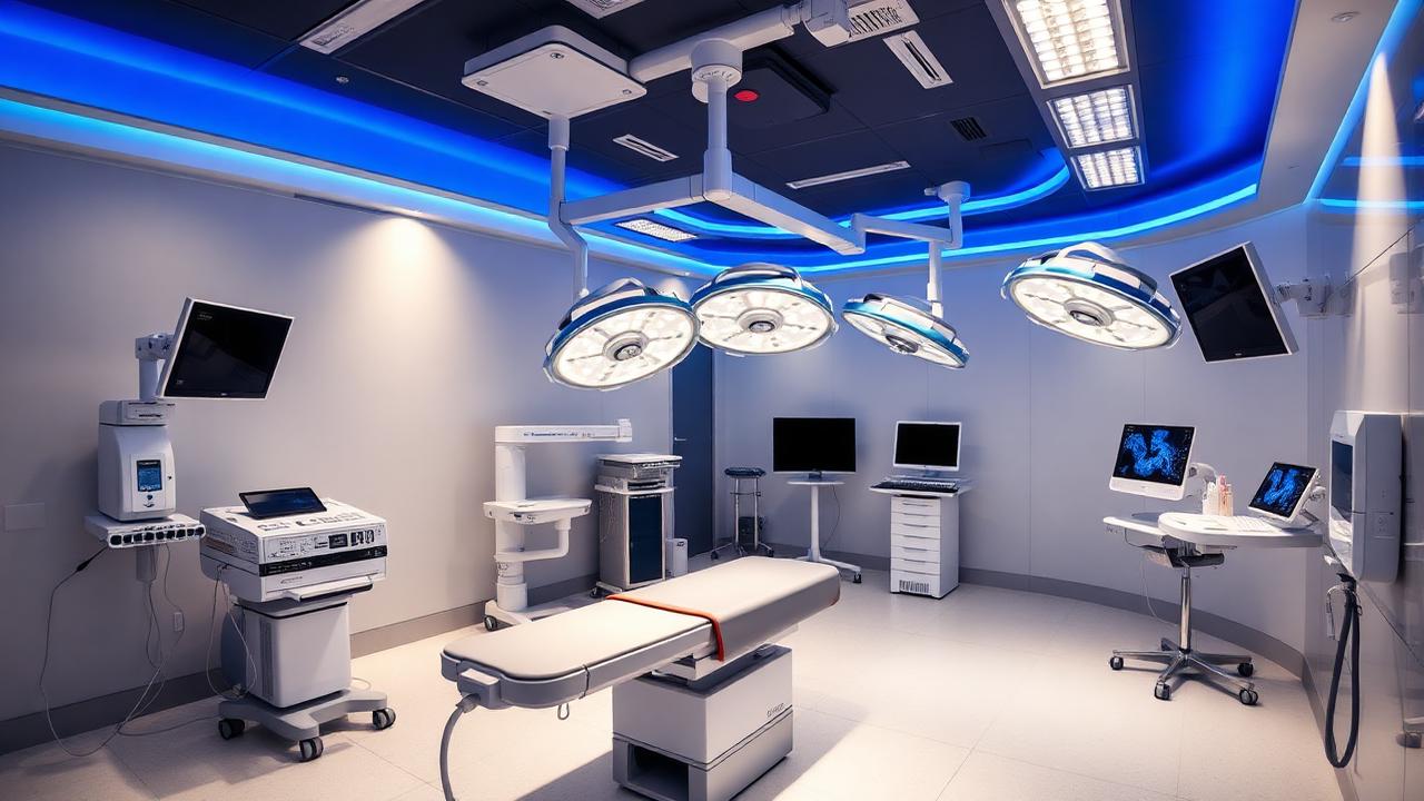
Ovarian Vein Embolization Explained, Step by Step
A detailed, procedure-day walkthrough of ovarian vein embolization for pelvic congestion syndrome, covering access, venography, coils, recovery, risks and realistic success rates.
Why Ovarian Vein Embolization Is Even Needed
Ovarian vein embolization is a minimally invasive, image-guided procedure used to treat pelvic congestion syndrome, a condition in which the ovarian veins and other pelvic veins become dilated and their one-way valves stop working properly, allowing blood to pool in the pelvis instead of returning efficiently to the heart.
This pooling raises pressure in the pelvic venous plexus, and over months or years that sustained pressure produces a dull, dragging ache that is typically worse by evening, worse after standing for long periods, worse premenstrually, and often improved by lying flat.
Because the symptoms overlap heavily with common gynaecological complaints, many women go through several rounds of scans and even diagnostic laparoscopy before a vascular cause is seriously considered, and by the time they reach a vascular surgeon the pain has often been present for years.
Who Is Actually a Candidate
Not every woman with pelvic pain has pelvic congestion syndrome, and not every case of pelvic congestion syndrome needs embolization. The typical candidate is a woman, usually of reproductive age and often with more than one prior pregnancy, who has chronic pelvic pain lasting more than six months, a normal or non-explanatory gynaecological examination, and imaging evidence of dilated, refluxing ovarian or pelvic veins.
Imaging usually starts with a transvaginal ultrasound with Doppler, which can show tortuous veins and reversed flow, and is often followed by a CT or MR venogram to map the full extent of the venous network, including the internal iliac and pelvic floor veins that a simple ultrasound may not fully capture.
A careful history is just as important as the scans. Dr. Pritee Sharma routinely asks about pain patterns tied to posture, the menstrual cycle, intercourse, and prolonged standing, because pelvic congestion syndrome has a fairly distinctive rhythm compared with, say, endometriosis or a urinary tract cause.
The Pre-Procedure Work-Up
Before the day of the procedure, a set of baseline blood tests is done, including kidney function and clotting parameters, since the procedure uses intravenous contrast dye and there is a small, controlled use of anticoagulation during the case.
Patients are counselled on what to expect, including the fact that this is done under local anaesthesia with sedation in most cases, that they will be awake but comfortable, and that the entire hospital stay is typically limited to a day-care admission with a few hours of monitoring afterward.
Any current medications, particularly blood thinners, are reviewed and adjusted in advance in consultation with the treating physician, and women are advised to fast for a short period before the procedure in case sedation needs to be deepened.
Admission on the Day of the Procedure
On the day itself, the patient is admitted to the day-care unit, changes into a hospital gown, and has an intravenous line placed in the arm for fluids, sedation, and any medication that might be needed during the case.
Vital signs are recorded and a final check is done to confirm consent, allergies, and the specific plan for that patient, since not everyone needs both ovarian veins treated and some also require additional embolization of pelvic floor veins.
Choosing the Access Point: Jugular or Femoral Vein
The procedure is performed by accessing the venous system through either the internal jugular vein in the neck or the femoral vein in the groin, and the choice depends on the specific venous anatomy, the angle needed to catheterize the ovarian veins, and operator preference.
Right internal jugular access is often favoured because it gives a straighter, more direct path down into both the left and right ovarian veins, which can make catheter manipulation faster and reduce total procedure time, while femoral access remains a very reasonable alternative and is sometimes preferred based on individual anatomy.
Whichever site is chosen, it is cleaned with antiseptic solution, numbed with local anaesthetic, and accessed using a fine needle and a sheath under ultrasound guidance, so the patient generally feels only a brief pinch rather than ongoing discomfort.
Venography: Mapping the Problem Veins
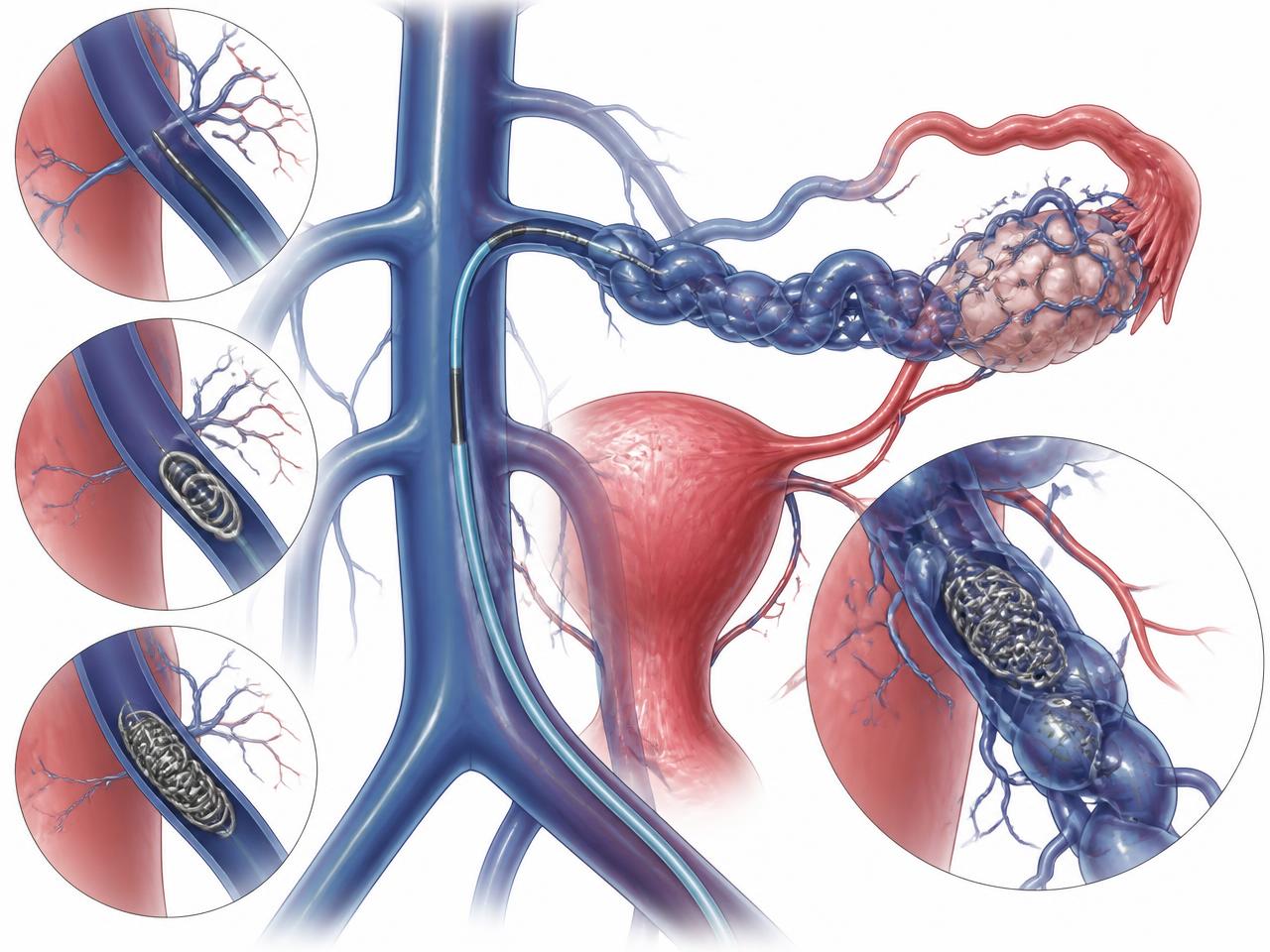
Once access is secured, a thin catheter is guided under x-ray (fluoroscopic) guidance into the left and then the right ovarian vein in turn. Contrast dye is injected and live x-ray images, called venograms, are taken to confirm the diagnosis in real time.
A competent ovarian vein normally shows contrast draining upward without much delay, whereas an incompetent vein in pelvic congestion syndrome typically shows dilation, tortuosity, and reflux of contrast down toward the pelvis, often filling a visible network of engorged pelvic and vulval veins.
This live mapping step is important because it tells the treating surgeon exactly which veins are contributing to the problem and in what order they should be treated, and it also rules out other causes such as nutcracker syndrome or May-Thurner syndrome, which sometimes coexist and may need a different treatment approach altogether.
Coils, Plugs and Sclerosant: How the Vein Is Closed
Once the abnormal vein is confirmed, it is treated by deploying tiny metal coils, and sometimes a combination of coils with a liquid or foam sclerosant, along the length of the vein from its lower end near the pelvis up toward its junction with the larger vein it drains into.
The coils are made of platinum or a similar biocompatible metal and act as a scaffold that triggers the body's natural clotting process, permanently sealing the vein so blood can no longer flow through it or reflux backward into the pelvis.
Sclerosant, when used, irritates the inner lining of the vein and causes it to scar shut, and is particularly useful for smaller tributary veins that coils alone might not fully address. The combination approach has become common practice because it reduces the chance of the vein reopening or of small collateral channels forming later.
Confirming the Result Before Finishing
After the coils are placed, a repeat venogram is done to confirm that contrast no longer flows through the treated vein and that blood flow has been satisfactorily rerouted. If a second or third vein needs treatment, for instance the opposite ovarian vein or an internal iliac tributary, the same steps of catheterization, venography, and embolization are repeated.
The whole procedure typically takes between forty-five minutes and two hours depending on how many veins need treatment and how tortuous the anatomy is, and it is done with the patient talking, breathing normally, and generally comfortable throughout.
Closing Up and Immediate Recovery
Once the venogram confirms a good result, the catheter and sheath are removed and firm pressure is applied to the access site for a few minutes to prevent bleeding, followed by a simple dressing. No stitches are usually needed since the access point is a small needle puncture rather than a surgical incision.
The patient is then moved to a recovery area where vital signs are monitored for a few hours. Mild pressure or ache at the access site and a dull pelvic cramp for a day or two are common and expected, and are managed with simple pain relief such as paracetamol.
Day-Care Discharge and What Happens That Evening
Most women are discharged the same evening once they are alert, able to walk, tolerating food and water, and have no signs of bleeding at the access site. A responsible adult is asked to accompany the patient home, since sedation can leave residual drowsiness for several hours.
Discharge instructions typically include keeping the puncture site dry for twenty-four hours, avoiding strenuous exercise and heavy lifting for about a week, and watching for warning signs such as spreading bruising, fever, worsening pain, or swelling at the access site that would warrant an urgent call to the hospital.
Risks and Complications to Understand Honestly
Ovarian vein embolization is generally very safe, but like any procedure it carries some risks that should be discussed openly rather than glossed over. Access site bruising or a small haematoma is the most common minor issue and usually resolves on its own within one to two weeks.
Coil migration, where a deployed coil moves from its intended position, is uncommon with modern deployment techniques and careful sizing, but is a recognised risk that is discussed during consent; if it occurs, it can usually be retrieved or managed without further surgery.
Other infrequent risks include an allergic reaction to the contrast dye, a small chance of infection at the access site, temporary flank or pelvic pain from the sclerosant, and in very rare cases, vein perforation during catheter manipulation. Radiation exposure from the fluoroscopy is kept as low as reasonably possible and is not considered a significant concern for this type of procedure.
- Bruising or tenderness at the neck or groin access site
- Temporary pelvic ache or cramping for a few days
- Rare allergic reaction to contrast dye
- Uncommon coil migration, usually manageable without repeat surgery
- Very rare vein injury during catheter placement
- Small risk of the treated vein or a collateral reopening over time
Success Rates: What Realistic Relief Looks Like
Across published clinical experience and routine practice, the majority of women who undergo ovarian vein embolization for well-diagnosed pelvic congestion syndrome report meaningful improvement in their pelvic pain, with commonly cited relief rates in the broad range of seventy-five to eighty-five percent at several months of follow-up.
Meaningful relief does not always mean the pain disappears completely from day one. Many women notice a gradual reduction in the dragging, heavy ache over the first four to six weeks as the treated veins scar down and pelvic venous pressure normalises, with continued improvement over the following months.
A smaller proportion of women have only partial relief, often because a coexisting cause such as endometriosis, a musculoskeletal pelvic floor issue, or an untreated additional vein is also contributing, which is why thorough pre-procedure imaging and honest expectation-setting matter so much.
Follow-Up After the Procedure
A follow-up visit is usually scheduled around two to four weeks after the procedure to check the access site, review symptom diaries, and address any residual discomfort. A follow-up ultrasound or venogram is occasionally used later if symptoms persist, to check whether the treated veins have remained closed or whether new collateral veins have developed.
Women are encouraged to keep a simple pain and activity diary in the weeks after the procedure, since this makes it much easier to have an objective conversation at follow-up about how much things have actually changed, rather than relying on memory alone.
Indicative Costs in Hyderabad and Insurance Notes
As an indicative range only, ovarian vein embolization in a Hyderabad private hospital setting, including day-care admission, catheters, coils, and physician fees, commonly falls somewhere in the region of INR 90,000 to INR 1,80,000, with the final figure depending on how many veins are treated, the number and type of coils used, and whether sclerosant is also required.
Many health insurance policies in India do cover this procedure when it is supported by clear imaging evidence and a documented diagnosis of pelvic congestion syndrome, but pre-authorisation requirements and covered amounts vary considerably between insurers, so it is worth getting a written cost estimate and checking policy-specific coverage before the day of the procedure.
Distinguishing Pelvic Congestion Pain From Gynaecological Pain
One of the more useful things a woman can do before her first vascular consultation is to notice the pattern of her pain rather than just its intensity. Pelvic congestion pain tends to build through the day, worsen with prolonged standing, ease when lying down, and often intensify in the days before a period, whereas pain from fibroids, endometriosis, or ovarian cysts tends to have a different rhythm tied more tightly to the menstrual cycle itself or to specific triggers like intercourse in a fixed position.
It is also common for pelvic congestion syndrome to be accompanied by visible signs elsewhere, such as varicose veins around the vulva, inner thigh, or buttock, or a feeling of heaviness that mirrors leg vein disease, which is one reason a vascular opinion is worth seeking even when gynaecological scans have come back unremarkable.
Red Flags That Need Urgent Review, Procedure or Not
Whether or not embolization has been done, certain symptoms should never be dismissed as ordinary pelvic congestion and need urgent medical review: sudden severe pelvic pain unlike the usual dragging ache, fever with pelvic pain, heavy or irregular vaginal bleeding, a palpable pelvic mass, or pain associated with fainting or significant blood loss.
After the procedure specifically, warning signs that warrant an immediate call to the treating hospital include spreading redness or swelling at the access site, fever above thirty-eight degrees Celsius, worsening rather than improving pain after the first week, or any sign of bleeding that does not stop with gentle pressure.
Bringing It All Together
Pelvic congestion syndrome remains under-recognised in general practice, partly because it does not show up on a standard pelvic ultrasound done for other reasons, a gap that Dr. Pritee Sharma discussed at length in her Times of India feature on pelvic congestion syndrome, where she highlighted how many women are labelled as having psychosomatic pain before the venous cause is even considered.
Ovarian vein embolization is a well-established, minimally invasive option for a condition that has historically been under-diagnosed and under-treated, and understanding the actual step-by-step flow of the procedure, from admission through access, venography, coil placement, and same-day discharge, often reduces much of the anxiety that comes with an unfamiliar intervention.
It is worth remembering that pelvic varicose veins and the more visible varicose veins of the leg often share the same underlying valve failure and hormonal drivers, and readers who also notice bulging veins on the thigh or calf can learn more about evaluation and treatment options at /services/varicose-veins-treatment-hyderabad.
If any of this sounds familiar, the Pelvic Vein Clinic at Renova Century Hospital, Banjara Hills, is set up specifically to evaluate and treat pelvic venous disorders under one roof, from the first ultrasound to the embolization procedure itself; you can read more about the clinic's approach and book a consultation at /pelvic-vein-clinic.
Dr. Pritee Sharma is a DNB Gold Medalist vascular and endovascular surgeon with over 21 years of experience; her background and areas of special interest are detailed at /about-dr-pritee-sharma, and questions about scheduling a consultation can be sent through /contact.
- Varicose Veins Treatment in Hyderabad
- Peripheral Artery Disease Treatment
- Diabetic Foot Care Specialist
- DVT Treatment in Hyderabad
- Angioplasty for Blocked Leg Arteries
- Carotid Surgery — Stroke Prevention
- Venous Stenting for Chronic Leg Swelling
- AV Fistula Creation for Dialysis
- EVAR — Endovascular Aneurysm Repair
- TEVAR — Thoracic Aortic Repair
- About Dr. Pritee Sharma
- Vascular surgery FAQs